Extensive brain activity while listening to speech suggests awareness in minimally conscious patients
By Ernie Mundell

NEW YORK (Feb. 7, 2005) -- For the first time, advanced neurological imaging suggests the brains of minimally conscious patients recognize and respond to speech in ways similar to healthy individuals, according to a team of researchers from Columbia University Medical Center and the Weill Medical College of Cornell University, both in New York City; and the JFK Johnson Rehabilitation Institute-Center for Head Injuries, in Edison, N.J.
"The results challenge our thinking about the possible inner lives these severely brain-damaged patients may experience, and also motivate renewed interest in research aimed at recovery and rehabilitation," said the study's senior author, Joy Hirsch, Ph.D., Professor of Neuroradiology and Psychology, and Director of the fMRI Research Center at Columbia University Medical Center.
The study is published in the February 8 issue of Neurology.
The minimally conscious state is distinct from either a persistent vegetative state or coma in that patients show intermittent signs of awareness and may even attempt to communicate using simple words or signals.
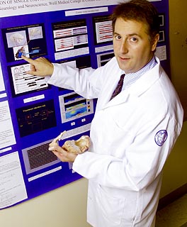
"Unfortunately, these episodes are erratic, so that most of the time caregivers and loved ones are left unable to make contact with the patient," explained study lead author Nicholas D. Schiff, M.D., Assistant Professor of Neurology and Neuroscience, and Director of the Laboratory of Cognitive Neuromodulation at Weill Cornell Medical College. Dr. Schiff is also Assistant Attending Neurologist at NewYork-Presbyterian Hospital/Weill Cornell Medical Center.
"In fact, many observers remain doubtful whether minimally conscious patients are even aware of the external world," he said.
To help shed light on that mystery, Dr. Schiff turned to the state-of-the-art functional magnetic resonance imaging (fMRI) at Dr. Hirsch's lab at Columbia. These studies, which have potentially broad implications for rehabilitation, arose from ongoing collaborations between Dr. Schiff and Joseph T. Giacino, Ph.D., Associate Director of Neuropsychology at the JFK Johnson Rehabilitation Institute.
Using fMRI -- which employs MRI scanning technology to track real-time brain function -- the team compared neurological activity in seven healthy volunteers with that of two minimally conscious patients, both young men. One of the minimally conscious subjects had experienced sudden brain hemorrhage, while the other's injuries were caused by blunt trauma to the head.
During the experiment, study subjects were exposed to audio of family members reading a simple narrative aloud, as researchers observed brain activity via fMRI.
"The results were a big surprise," Dr. Schiff said. "The spoken narratives appeared to activate language networks across many different regions of the brain, in both normal and brain-damaged subjects. In fact, in at least one of the minimally conscious patients we studied, all of the components that were active in healthy subjects were also active in his brain."
A second finding the researchers described as "haunting" suggests an even more personal response to speech on the part of minimally conscious subjects.
"Especially in one of the two patients, the spoken narratives appeared to activate visual areas of the brain," Dr. Hirsch said. "This happens in healthy subjects whenever language evokes visual images."
In a less encouraging finding, the researchers say the "resting activity rate" of the brains of minimally conscious patients remains very low, suggesting they lack the metabolic resources to keep specific cognitive networks running at a consistently high level. The finding could help explain the intermittent, episodic nature of awareness and communication seen in minimally conscious patients.
"Obviously, we still need to find out so much more about the neurological potential of these patients, so research building on these findings is already underway" Dr. Schiff said.
Technology is at the very heart of this research, Dr. Hirsch added. "Brain imaging can be thought of as a way of giving a real voice to the minimally conscious patient," she explained. "It's allowing us as physicians and scientists to be more aware of these patients' potential for rehabilitation."
For his part, Dr. Giacino called the findings "exciting news."
"While it's far too early to speculate as to therapies, those of us working in the rehabilitation sphere now have real evidence these severely damaged brains retain essential networks," he said. "These studies should encourage further efforts at rehabilitation to try and break these patients out of their isolation, to better understand what their capacity to experience -- and interact with -- their environment really is," he added.
This research was supported with funding from the National Cancer Institute, the Charles A. Dana Foundation, the National Institute of Neurological Disorders and Stroke, and the NIH-supported NewYork-Presbyterian/Weill Cornell General Clinical Research Center.
Co-researchers include Dr. Fred Plum and Dr. A. Kamal of Weill Cornell Medical College; Diana Rodriguez-Moreno of Weill CornellÕs Graduate School of Medical Sciences; and Dr. K.H.S. Kim of Georgetown University Medical Center (Washington, D.C.).
Ernie Mundell is a writer at Weill Cornell.
Media Contact
Get Cornell news delivered right to your inbox.
Subscribe