Viruses discovered for first time in marine zooplankton
By Krishna Ramanujan

Viruses are well known for making people sick, but a new study provides evidence for the first time of viral infections in tiny marine crustaceans called copepods.
While predation by fish and other aquatic creatures accounts for the majority of copepod deaths, up to 35 percent of the zooplankton's (tiny organisms) mortalities are unknown. Harmful algae, environmental stressors, parasites and diseases are likely all involved in copepod mortalities.
A study published in the Jan. 7 issue of the Proceedings of the National Academies of Science uses genomic techniques to make a compelling case for viral infections as a major cause of copepod deaths.
"This is the first evidence of viruses in marine zooplankton," said Ian Hewson, Cornell assistant professor of microbiology and senior author of the paper. Darren Dunlap, a graduate student at the University of South Florida, is the paper's lead author.
The finding is important because copepods are critical in oceanic food webs and ocean carbon cycling, a key process for regulating Earth's climate. Marine free-floating plants called phytoplankton sequester about half of the carbon dioxide that is pulled from the atmosphere and fixed in plant cells during photosynthesis. Copepods play a key role in the process by consuming most of the oceans' phytoplankton, which they defecate as pellets that sink to the ocean floor, thereby locking up atmospheric carbon for thousands of years. Quantifying these dynamics is important as carbon dioxide, a major greenhouse gas, is increasing in the atmosphere due to human activities and is changing Earth's climate, according to the Intergovernmental Panel on Climate Change.
"We now have identified another aspect of the microbial food web that affects the amount of atmospheric carbon that potentially gets fixed," Hewson said. Such information will be important to researchers who use computer models to understand population dynamics, food webs and climate change.
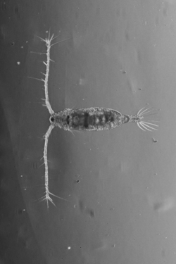
The researchers used microscopic and molecular techniques to document viral infections in natural populations of two common copepods, Acartia tonsa, a globally distributed herbivore, commonly raised for fish meal, and Labidocera aestiva, a large omnivore that is important in estuary ecosystems.
Using a genomic technique, Hewson, Dunlap and colleagues removed viruses found in the copepod tissues, sheared their DNA and identified known viral DNA sequences. They pinpointed two previously undocumented viruses, each unique to L. aestiva and L. tonsa, respectively. Both viruses belong to the Circovirus group, which has previously only been observed in pigs and birds, and more recently in some other arthropods, including flying insects. These copepod-specific viruses were not found in any other zooplankton species.
"Genomics allows us to identify something we otherwise might have missed using other techniques," Hewson said.
Infection rates varied by population and season, with average viral loads of hundreds of thousands of viruses per copepod and 100 percent of individuals infected in some communities of L. aestiva. Though longer term data is needed, the findings suggest that the virus that infects L. tonsa may rise in the spring and autumn and drop in the summer and winter as copepod populations rise and fall.
Using electron microscopy, the researchers identified virus-like structures in copepod tissues, showing these viruses were not infecting the guts or associated with gut parasites that may carry them.
Future work will evaluate the viruses' pathology and mortality rates on copepods, route of infection and ecological implications.
The study was funded by the National Science Foundation.
Media Contact
Get Cornell news delivered right to your inbox.
Subscribe